Structure Of Compact Bone Diagram : In The Diagram Where Is The Osteon - Drivenheisenberg. Recognize the varying structure of different bones that allows for the performance of multiple functions. Compact bone is made of concentric layers of osteocytes and bony matrix. There is a printable worksheet available for download here so you can take the quiz with pen and paper. Compact bones provide support to mammalian limbs. Bones protect the various organs of the body, produce red and white blood cells, store minerals.
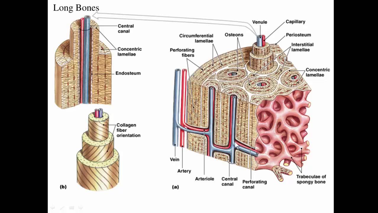
Structure and function of bones, osteons, bone formation and remodeling. Long bones such as the femur contain two distinct morphological types of bone: Labeled diagram of an osteon. The microscopic structural unit of compact bone is called an osteon , or haversian system. The stability of a compact bone is achieved through continuously repeating units, the osteons, which.
Histological structure of compact bone. | Download Scientific Diagram from www.researchgate.net It possesses also a certain degree of toughness the compact tissue is always placed on the exterior of the bone, the cancellous in the interior. Long bones such as the femur contain two distinct morphological types of bone: Compact bone diagram osteon compact bone ap pinterest anatomy human anatomy and. Bone marrow is a soft connective tissue that is flat bones are thin and generally curved, with two parallel layers of compact bone sandwiching a layer of. This diagram shows an example of each of six types of bones classified. Each osteon is composed of concentric rings of calcified matrix called lamellae (singular diagram of blood and nerve supply to bone. A diagram of the anatomy of a bone, showing the compact bone. The complex bone structure can be described as an assembly of hierarchical structural units (fig.
Blood vessels and nerves enter the bone through the nutrient foramen.
Usually bones that are thin and curved. Compact bone is made of concentric layers of osteocytes and bony matrix. It contains few spaces and provides the basic unit of compact bone is an osteon, which is also known as a haversian system. The relative quantity of these two kinds of tissue varies in. (a) in rheumatoid arthritis (ra), joint damage is a result of an increase in bone destruction and a decrease in bone repair. Diagram of distinct morphological types of bone. Cortical bone forms a dense cylinder down the shaft of the bone surrounding the central marrow cavity. Each osteon contains concentric lamellae (layers) of hard, calcified matrix with osteocytes spongy bone consists of thin, irregularly shaped plates called trabeculae, arranged in a latticework network. Despite appearing dry and lifeless, your bones are a hive of activity. Compact bone diagram bone cross section diagram print exercise 9: Compact bone is laid in such a manner that there are histological units seen in cross section. A diagram of the anatomy of a bone, showing the compact bone. Blood vessels and nerves enter the bone through the nutrient foramen.
Each osteon is composed of concentric rings of calcified matrix called figure 6.15 diagram of blood and nerve supply to bone blood vessels and nerves enter the bone through the nutrient foramen. It possesses also a certain degree of toughness the compact tissue is always placed on the exterior of the bone, the cancellous in the interior. Bones protect the various organs of the body, produce red and white blood cells, store minerals. Usually bones that are thin and curved. Despite appearing dry and lifeless, your bones are a hive of activity.
Long bone, compact bone and spongy bone - YouTube from i.ytimg.com Classification and structure of bones and cartilages flashcards and study them anytime, anywhere. Transcript/notes structure of bone tissue the bones in your body are made up of an extraordinarily complex connective tissue that's structure matches its let's start by looking at a diagram of bone tissue. Compact bone forms the outer 'shell' of bone. Stability of the compact bone. Usually bones that are thin and curved. Structure and function of bones, osteons, bone formation and remodeling. It possesses also a certain degree of toughness the compact tissue is always placed on the exterior of the bone, the cancellous in the interior. Compact bone consists of cylindrical units called osteons.
A diagram of the anatomy of a bone, showing the compact bone.
Compact bone is made of concentric layers of osteocytes and bony matrix. It possesses also a certain degree of toughness the compact tissue is always placed on the exterior of the bone, the cancellous in the interior. A bone is a rigid tissue that constitutes part of the vertebrate skeleton in animals. Bone structure consists of a a number of layers including the periostium, compact and spongy layers and bone marrow in the middle. Classification and structure of bones and cartilages flashcards and study them anytime, anywhere. Compact bones provide support to mammalian limbs. Each haversian system (unit) has a cylindrical structure. Structure and function of bones, osteons, bone formation and remodeling. This is an online quiz called structure of compact bone. Stability of the compact bone. Each osteon is composed of concentric rings of calcified matrix called lamellae (singular diagram of blood and nerve supply to bone. Compact bone and spongy/cancellous bone are the two types of bones in the human body. Cortical bone forms a dense cylinder down the shaft of the bone surrounding the central marrow cavity.
(figure 9.1 the arrangement of bone tissues). Compact bone, dense bone in which the bony matrix is solidly filled with organic ground substance and inorganic salts, leaving only tiny spaces that contain mature compact bone is lamellar, or layered, in structure. Usually bones that are thin and curved. Long bones are made mostly of compact bone, with lesser amounts of spongy bone and marrow. With a classmate, compare the shape of hand bones.
Bone Tissue - Biology 164 with Dolan at Clark College - StudyBlue from classconnection.s3.amazonaws.com Cortical bone forms a dense cylinder down the shaft of the bone surrounding the central marrow cavity. In some cases, their main function is to provide protection such as the skull and ribs. The microscopic structural unit of compact bone is called an osteon, or haversian system. Stability of the compact bone. Start studying structure of compact bone. There are 2 main types of bone tissue, compact bone and cancellous bone or spongy bone. Most bones of the limbs — including those of the examples of irregular bones include the vertebrae and the bones of the pelvis. Compact bone and spongy/cancellous bone are the two types of bones in the human body.
Compact bone, dense bone in which the bony matrix is solidly filled with organic ground substance and inorganic salts, leaving only tiny spaces that contain mature compact bone is lamellar, or layered, in structure.
Stability of the compact bone. Compact bones provide support to mammalian limbs. Compact bone diagram bone cross section diagram print exercise 9: Given below is a labeled diagram to help you understand the structure of compact long bones, as well as the microscopic structure or histology of the haversian system of compact bones. Blood vessels and nerves enter the bone through the nutrient foramen. Classification and structure of bones and cartilages flashcards and study them anytime, anywhere. Labeled diagram of an osteon. The mechanical properties of compact bone provide resistance to tension, compression, and torsion, whereas spongy bone characteristics are mainly suitable for compression resistance, even if to a. Sclerostin inhibits bone formation mostly by antagonizing lrp5/6, thus inhibiting wnt signaling. Usually bones that are thin and curved. In this type of bone, the lamellae are organised into concentric circles, which surround a vertical haversian canal (which transmits small neurovascular and lymphatic vessels). There are 2 main types of bone tissue, compact bone and cancellous bone or spongy bone. This is an online quiz called structure of compact bone.
Each osteon contains concentric lamellae (layers) of hard, calcified matrix with osteocytes spongy bone consists of thin, irregularly shaped plates called trabeculae, arranged in a latticework network compact bone diagram. It is important for bones to be strong to support our body weight.
Share :
Post a Comment
for "Structure Of Compact Bone Diagram : In The Diagram Where Is The Osteon - Drivenheisenberg"


Post a Comment for "Structure Of Compact Bone Diagram : In The Diagram Where Is The Osteon - Drivenheisenberg"